-
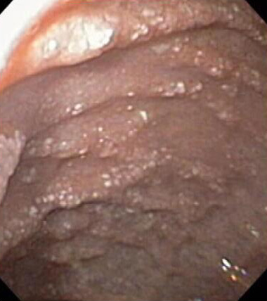
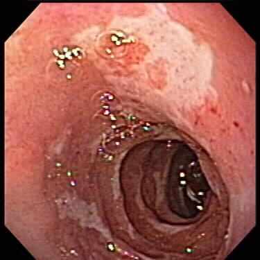
Intestinal Lymphangiectasia
Endcoscopic photograph of the 3rd portion of the duodenum in a 15 yo who presented with abdominal pain and diarrhea.
-
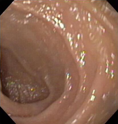
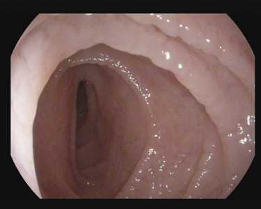
Normal Duodenum
Endoscopic photograph of a normal duodenum.
-

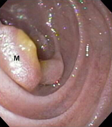
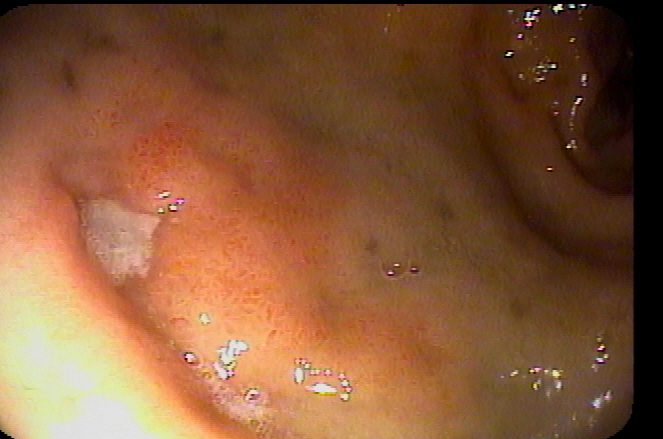
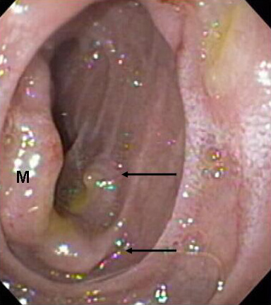
Duodenal PTLD
Endoscopic photograph of one large (M) and several small (arrows) masses visualized in a patient with vomiting and abdominal pain who previously had a renal transplant.
-
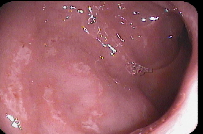
Duodenal PTLD
Endoscopic photograph of a large mass (M) visualized in a patient with vomiting and abdominal pain who previously had a renal transplant.
-
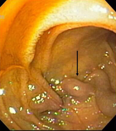
Duodenal Varix
Endoscopic photograph of a duodenal varix (arrow) in a 12 yo patient with portal hypertension. (CAL)
-
Duodenal ulcers (NSAIDs)
4 yo male with long history of NSAID exposure secondary to recurrent otitis who presented with abdominal pain and hematemesis.
-

NSAID induced ulcers
NSAID induced ulcers
-
NSAID induced ulcers
NSAID induced ulcers
-
Duodenal ulcer secondary to H pylori
Duodenal ulcer secondary to H pylori
-

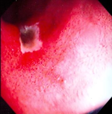
Duodenal ulcer
Duodenal ulcer. Endoscopic view of an ulcer in the duodenal bulb of a 12-year-old who presented with hematemesis.
-
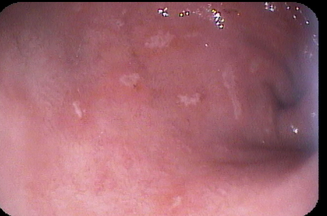
Celiac Disease
11 y/o with positive tissue transglutaminase (TTG). Multiple images demonstrating typical appearance of celiac disease. Image 4 enhanced with digital filter.
-

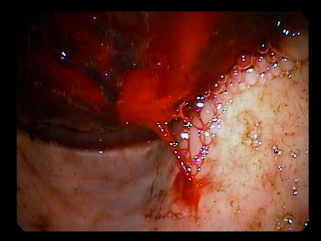
Portal Vein Occlusion
Portal vein occlusion post liver transplant causing incresed pressure gradient and bleeding at biliary-enteric anastomotic orifice
-
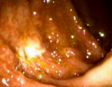
Duodenal Ulcer
Duodenal ulcer with fresh bleeding (Image 1), after multipolar electrocoagulation (Image 2)
Photos 1-13 (of 13)


.JPG)


Photos 1-13 (of 13)